Apoptosis: Programmed Cell Death — induced
Purpose:
— in cell development, some cells need to be killed in order to achieve the final structure
e.g. limb formation: Sonic Hedgehog allows the digits to be positioned, then apoptosis will kill the cells b/w the limbs to separate them
— kill of dangerous cells that can cause abnormalities such as cancer (recognize mistakes and allows it to be repaired)
e.g. Excessive Myc (drives cell proliferation) production can lead to cancer. If p53 cannot stop cell cycle or if the cell cannot mix itself then the prolonged signal of active p53 will activate apoptosis.
Necrosis: Accidental Cell Death, can lead to a damaging inflammatory reaction when the cell bursts. When the extracellular environment is contaminated by intracellular material, the neighbouring reaction is inflammation.
In contrast, apoptosis is neat and never disrupts other cells’ normal functions. It does this through breaking down the macromolecules inside the cell:
— cell shrinks
— cellular cytoskeleton collapses
— DNA is fragmented
Finally, the apoptotic cell (with all the interior components fragmented but still contained by the membrane) will signal to macrophages to be engulfed. This way the intracellular material never exposes to the external environment.
Molecular basis for triggering apoptosis
caspases:
- are proteases that cleave many downstream target proteins at specific aspartic acid residues
- synthesized as procaspase precursor that are always present but inactive in our bodies—> cleaved and activated by other caspases —> cascade activity
*The activation of a single caspase begins with two inactive procaspases that are then cleaved at 2 sites. Once cleaved, the prodomains on the NH2 end will be released, and the COOH ends go through a complex rearrangement so that 2 procaspases will come together to form a tetramer: active caspase.
* A small group of active initiator caspases can then activate many more caspases called executioner caspases: amplification cascade. These executioner caspases act on the intracellular proteins to break them down. Specific executioner caspases target specific proteins, not every cytosol proteins
Apoptosis progress can be detected:
by electrophoresis —> separating the DNA fragments after apoptosis induction.
by TUNEL —> transfer nucleotide into the termini to label them (the more DNA fragments, the more ends there will be, and brighter the signal)
by antibody that labeled activated caspases
by altered cell membrane —> in normal cells, the phospholipid phosphatidylserine is found only on the cytosolic side, but when the control mechanism for it breaks down due to apoptosis, the phospholipid will flip to the extracellular side, and can be detected by macrophages in vivo, and probes in vitro.
Activation of apoptosis
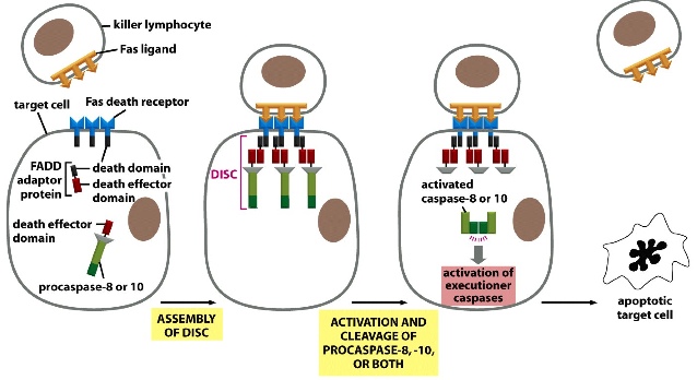
1) triggered by extrinsic signals via death receptors
e.g. killer lymphocyte will trigger its own apoptosis when it become infected by a virus so that it will limit danger to other cells to a minimum.
*FADD: Fas associated death domain
**DISC: Death inducing signalling complex
***Fas ligand expressed on the surface of killer lymphocyte + Fas death receptors expressed on the target cell (to be destroyed) —> draws in FADD adaptor protein —> links the receptor to procaspase (which has death effector domain) —> procaspase that have low activity when alone is able to cleave each other when they are drawn into close proximity —> activate executioner caspases —>cleave target components
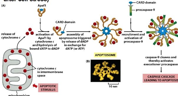
2) triggered by intrinsic signals from mitochondria
Electron carrier protein c (cytochrome c) is released from mitochondria under stress or other apoptotic simuli —> Cytochrome c binds to Apaf1 in the cytosol —> exposes the CARD domain of Apaf1 —> multiple singular structures then assemble into a large apoptosome —> recruits procaspase-CARD complex—> clusters them and bring them to proximity so that they can start cleaving and activating one another —> active initiator caspases then can activate executioner caspases
Here, apoptosis activity can be detected by tracing the location of stained cytochrome c in the cell —> mitochondria when normal; cytoplasm when apoptotic.
Extrinsic or intrinsic signals(cytochrome c) lead to the aggregation of top caspases promoting self-cleavage
Inhibition of apoptosis
Extracellular inhibitors
They are needed so that cells won’t commit suicide whenever they sense a signal even if it’s weak —> increasing the threshold of apoptosis activation by using inhibitors called decoy receptors on the surface of target cells:
The radio of decoy receptor: Fas death receptor can be altered according to the needs of the cells. There are always some Fas death receptors on the surface but usually not enough to trigger apoptosis. Decoy receptors increase the activation threshold by competing with Fas death receptors for Fas ligand binding. If the cell is infected by virus and needs to be destroyed, the cell will express many more Fas death receptors so there is a higher chance for Fas ligand to bind to Fas death receptor and initiates apoptosis.
Intracellular inhibitors
e.g. FLIP mimics an initiator caspase but cannot activate downstream degradation. FLIP doesn’t have death effector domain. This way although it competes with normal caspase to bind to upstream complexes, it will have no effect on proteins.
Other inhibitors can block apoptotic machinery
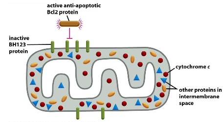
e.g. Bcl2
Bcl2 stops channel formation (formed by active BH123 proteins) on the surface of mitochondria so cytochrome c won’t be released through the channel
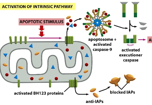
e.g. IAP (inhibitors of apoptosis)
bind to activated caspase tetramer in the cytosol. Constantly being made in the background so that it can prevent accidentally activated caspases to destroy cells at random.
Therefore, apoptosis requires active cytochrome c release and anti-IAPs.
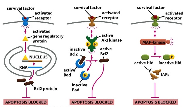
Such inhibitors can be regulated by survival factors
1) survival factors bind to surface receptors to activate gene regulatory proteins which then lead to the synthesis of Bcl2 proteins —> blocks apoptosis
2) survival factors bind to surface receptors to activate Akt kinases which releases Bad by phosphorylating it. This frees on Bcl2 formerly bound by Bad so that it can block apoptosis
3) survival factors bind to surface receptors to activate MAP kinase —> releases Hid by phosporylation —> active IAPs then blocks apoptosis
Regulation and maintenance of cell numbers
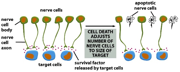
e.g. neuronal cell numbers is determined by competition for survival factors made by target cells
During brain development there are many axonal connections they we don’t need later on. Axons with the strongest connections receive survival factors and survive while other axons go through apoptosis.
Other tissues also regulate their own size through a combination of apoptosis and cell proliferation. Such as the case when the size of a liver is surgically altered, it uses apoptosis if it’s larger than normal and uses cell proliferation if it’s smaller than normal OR if it senses environmental stress.